Get Muscles Of The Thigh Mri Images. Piriformis, gemellus superior, obturator internus, gemellus inferior, obturator externus, and quadratus femoris main function: Along the posterior portion of the muscle (yellow arrows), there is a flat area of tendon originating from the knee.
Mri Of The Thigh Detailed Anatomy Superior Part W Radiology from w-radiology.com Thigh muscle strains are common for people of all ages. Muscles that move the leg are located in the thigh region. External rotation and abduction of the thigh;
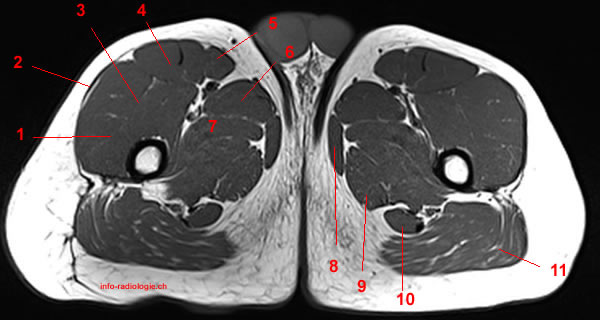
A magnetic resonance imaging (mri) was performed on a healthy subject;
The term 'hamstring' came about as it was common to use the long tendons of these muscles to tie up hams or pork thighs for curing. Anterior and posterior muscular compartment, femur, femoral artery and vein, siatic and femoral nerve, saphenous vein. The quadriceps femoris muscle group straightens the leg at the knee. Thigh muscles are responsible for allowing normal gait and it involves magnetic fields and radio waves to develop images of the body's internal organs(7).